Steve Grass,
the Technical Advisor and Trainer for Staub Cranial at
Select Dental Supply introduces the technology behind
Staub Cranial and explains the process. In this
introduction Mr. Grass explains how Staub Cranial is
able to "See the end from the beginning".
See the end from the
beginning
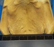
With Staub CranialRecently a new technology was introduced to the United States that will revolutionize the way our industry restores teeth. TheStaub Cranialsystem is a simple but effective way to locate the natural tooth position. Restorations are aligned based on cranial reference points and designed on the basis of mathematical calculations. This new approach challenges the use of average values or practitioner judgment and will set new standards both clinically and in the lab. There are times when restorative appliances have little if any resemblance to the natural dentition and have not been manufactured to be patient specific. This is especially true for denture wearers who commonly have two or three sets; one for looks, one for eating, etc. Dental technicians seldom see the patient and are left to manufacture what they believe a restoration should look like. The position, function, shape, and inclination are left to artistic discretion, not exact calculation. The premise of this technology is; every person has one (and only one) correct occlusal position. This leaves us with the question: Are we capable of calculating exact tooth placement? With Staub Cranial the answer is yes. Early in his career Master technician* Karl Heinz Staub (Neu- Ulm, Germany) asked the question, Why is the placement of teeth so subjective? Over the next twenty five years Mr. Staub researched and developed mathematical theorems that determine the exact position of the natural dentition. The results of this research led to the development of the Staub Cranial System . The University of Freiberg conducted a scientific validation of this system in the year 2003*. Two maxillary master casts were manufactured from twenty dentulous subjects. All teeth were removed on one of the two models made. Master technician Karl Heinz Staub, who was unaware of the previous tooth position, replaced the teeth for each of the twenty edentulous models. The replacement of the dental status deviated by only 5% of the original control models. (Figure 1) In other words Mr. Staub was able to replace the teeth with 95% accuracy.
  Figure 1
Today, Karl Heinz Staub and the Staub Cranial system are on the cutting edge of dental restorative technology. Many laboratories throughout Europe have adopted this exciting new technology. It was first introduced in the United States and Canada in February of 2007 and has quickly gained recognition. Staub Cranial utilizes definitive (bone) reference points to determine tooth placement. These points are anatomically stable and always present whether the patient is dentulous or edentulous. On the maxilla these locations are in the Hamular notch area, the Incisal papilla, and the free gingival attachment located on the labial portion of the alveolar process. The mandible has similar reference points (Figure 2). Staub identifies these as Direction points, Induction points, and Conclusion lines. 
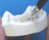
Figure 2 In his study Mr. Staub found that measurments between points (A, B, C and C1) created isosceles triangles thus making it possible to mathematically calculate the exact dental position. (Figures 3) 
 Figure 3 Using these points of reference Staub obtained mathematical theorems that allowed exact mapping of the ideal natural dentition (Figure 4).  Instruments created
by Mr. Staub are keyed to these measurements and allow
the articulation of the
maxillary model without the aide of a centric bite
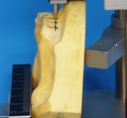
registration (Figure 5).  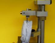
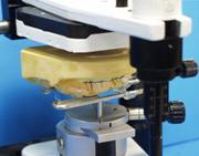 Figure 5 The correct placement of dentition on the maxilla has always been the keystone of dental restoration because of its definitive and conclusive reference points. Centric position can and should be calculable if we identify anatomically stable reference points on the maxilla and align the mandible accordingly. Once proper position is obtained restoration can then proceed. As an accurate assessment is performed by the dentist and then confirmed by the technician the accuracy of the final restoration is assured. Only a mathematical and scientific approach can guarantee this kind of consistent accuracy. The impressions are taken using special methods to capture these reference points. These points are then located on the working model by the technician, recorded, and measured. Accurate mounting of the maxilla is performed and a diagram of the proper dental placement is secured to an occlusal platform called the Staub Penta Plane. (Figure 6) 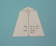 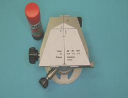 Figure 6 This technique is very applicable when evaluating full mouth cosmetic cases. For example: 1. Diagnostic wax-ups are evaluated and positioned to the patients ideal natural tooth position. 2. T.M.J. disorder patients now have a defined starting point for fabrication of a treatment splint. 3. Complex implant cases are simplified with the fabrication of predictable, precision surgical drill guides. These are manufactured with the clear understanding of the appropriate physiologic parameters of the final restoration. 4. Ceramic substructures are designed with definitive understanding of the location of the final porcelain restoration. The process is simple and easy to learn. A model map is produced by measuring from A, B, then B, C and B, C1. (Figure 7) These three measurements are entered into the computer software and a model map is produced. The model map is cut out and glued to the penta plane on the Ortho 3A. (Figure 8)  Figure 7 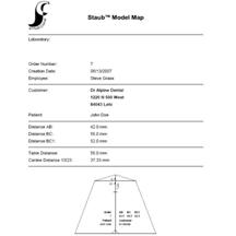
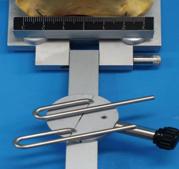
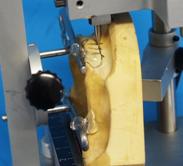
Figure 8 The maxillary model is positioned on the Ortho 1A according to the direction points, induction points, and conclusion lines. Proper positioning is of utmost importance and must be done precisely. This determines the exact position the maxillary model will be placed on the articulator. The Ortho 2A adapter and loops are positioned on the Ortho1A beam and the loops are position and secured to the maxillary ridge. (Figure 9)
Figure 9
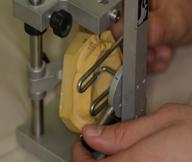
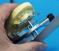
The attached model and adapter are removed from the beam and placed back on the Ortho 2A base. The Ortho 2A is placed on the articulator base and the maxillary model is attached to the articulator with plaster. (Figure 10) 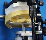
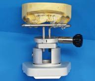 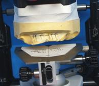 Figure 10 The Ortho 2A is removed from the articulator and replaced with the Ortho 3A. Anterior and posterior teeth are positioned in clay on the model map, the base plate is manufactured, and the teeth are attached to the model and base plate with wax. Wax and carving of the gingival is now performed and the upper wax up is complete. (Figure 11) 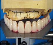
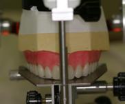 Figure 11 The mandible is mounted and evaluated in much the same way as the maxilla. The lower anterior teeth are set and posterior bite blocks are manufactured. The lower posterior bite block will aide in refining the bite relationship at the try in. (Figure 12) 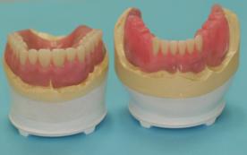
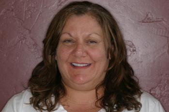
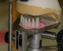 Figure 12 The case is sent to the doctor for try in. After the doctor evaluates aesthetic considerations a bite registration is obtained. The case is returned to the lab for final adjustments, and processing. Instead of five or six patient appointments only three are needed. The patient receives a restoration that is scientifically designed for His or Her mouth. The following case study was accomplished using the Staub Cranial system. The Patient desires to have a more natural smile and would like to have the ability to chew food. She feels her teeth are slanted and wants straight white teeth similar to what her teeth looked like when she was younger.(Figure 13) She provided a photograph of when she was a teenager so that her new set of teeth would have a similar look. The master casts are produced and measured according to the Staub Cranial method and preparations are made to set the teeth. The patient is included in the selection of the tooth shade and the upper denture is set for a try in. 
Figure 13 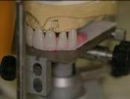 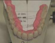
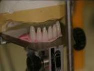 Figure 14 The lower anterior teeth are set and posterior bite blocks are manufactured. This entire procedure up to this point has been accomplished without the aide of a bite registration. (Figure 14) The case is now sent to the doctor for try-in. The patient is ecstatic with the results and the case is prepared to finish. The processed case is now ready for final placement. (Figure 15) 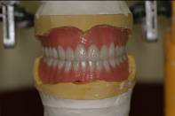
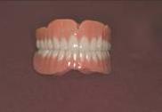 Figure 15
 The Staub Cranial final restoration is both aesthetically pleasing and functionally accurate. The Doctor and the Technician have worked in perfect harmony and understanding using scientific and mathematical accuracy. Guesswork and personal preference have not interfered. As the patient in our example left the office she threw her old dentures into the garage. We tried to convince her that should keep them as a spare but she said she was so happy with her new dentures that she never wanted to wear the old ones again. The dental industry has always attracted highly skilled Doctors and Technicians. Although artistry is and always has been a part of our industry, we must remember that patient specific placement of the dental arch should only be assessed through scientific and mathematical means. In a world where aesthetics has replaced function and fit as the number one consideration, we must be careful not to compromise proper dental placement. Many laboratories throughout Europe are now using this technique, and it has received great interest since its initial introduction to the North American continent in 2007. This scientific and mathematical approach will truly change the way we do dentistry and set a precedent in dental procedures. We will be able to accurately determine and see the end before we begin.  *Karl Heinz Staub of
Neu ulm, Germany is the founder of the Staub Cranial
system. He is a master technician and has devoted over
25 years to study and research of this exciting new
technology. *Karl Heinz Staub of
Neu ulm, Germany is the founder of the Staub Cranial
system. He is a master technician and has devoted over
25 years to study and research of this exciting new
technology. | |||||||||||
Blending Art & Technology
Wildomar CA 800-300-7614
$100.00 off your first Staub Cranial, Premium Denture
or Partial (HERE)
Staub Cranial Computerized Denture System "In Depth"
Nothing new for the Dentist to Buy or Learn!!
|
Download Newsletter
 | |||||||||||